



MRI (Magnetic Resonance Imaging)
Magnetic Resonance Imaging (MRI) is useful for creating detailed images of the inside of the body, particularly soft tissues like the brain, spinal cord, and joints that are not covered by bone. This imaging is especially performed on joints and bones, including the spine and limbs.
MRI imaging at the Iran Rheumatism Center is available in an open configuration for patients with claustrophobia, as well as in a standing position at this center.
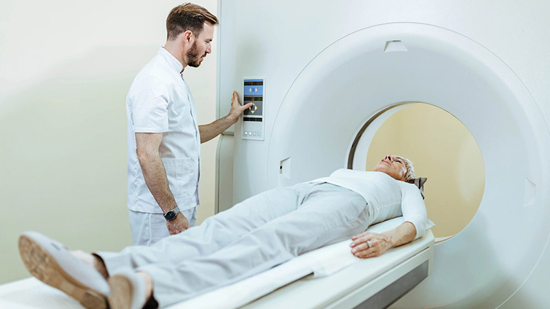
Frequently Asked Questions about MRI
MRI (Magnetic Resonance Imaging) is an advanced imaging technique that uses a strong magnetic field and radio waves to create detailed images of internal body organs (such as the brain, spinal cord, joints, and abdominal organs). It is used to diagnose conditions like tumors, nerve damage, inflammation, or ligament tears.
No, MRI does not use X-rays (ionizing radiation) and is safe for most people. However, if you have implanted metal devices (such as a pacemaker, certain stents, or shrapnel), you must inform your doctor before the test.
- Wear metal-free clothing (e.g., a hospital gown).
- Remove all jewelry, watches, and metal objects.
- Fasting may be required if a contrast agent will be used.
- Inform your doctor if you are pregnant or have kidney disease.
No, but the MRI machine is a closed tunnel, which may be uncomfortable for people with claustrophobia. In such cases, your doctor may prescribe a sedative.
The knocking or thumping sounds occur due to the activation of the machine’s coils and radio waves. Earplugs or headphones are usually provided to minimize discomfort.
The procedure takes between 20 to 60 minutes (depending on the area being examined). You must remain still during the scan to ensure clear images.
MRI can be performed during pregnancy if necessary (especially after the first trimester). However, the use of contrast material (gadolinium) is generally not recommended during pregnancy.
MRI uses a magnetic field and is more precise for examining soft tissues (e.g., the brain or ligaments). CT scans, however, employ X-rays, are faster, and are better suited for imaging bones or internal bleeding.
Contrast material (usually gadolinium) is injected intravenously to enhance image detail. Side effects are rare but may include itching, headache, or nausea. Patients with kidney failure should exercise caution.
Images are viewable immediately after the scan, but the final interpretation by a radiologist may take 1 to 3 business days.